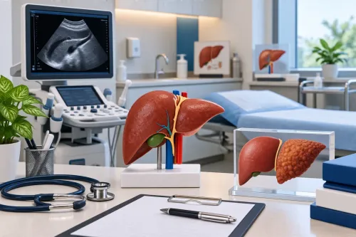
A liver biopsy has traditionally been an important method for examining liver tissue and assessing fibrosis. But many patients understandably ask whether there are liver biopsy alternatives for fibrosis assessment.
Today, non-invasive testing has become an important part of hepatology. Depending on the disease and clinical situation, blood-based calculations and imaging-based methods may provide useful information without requiring a tissue sample.
The important point is that these tests are not interchangeable in every patient. Choosing the right method depends on the suspected liver disease, fibrosis risk, body characteristics, previous test results, and diagnostic uncertainty.
What is fibrosis?
Fibrosis develops when repeated liver injury causes scar tissue to accumulate.
It can occur in conditions such as:
- Metabolic dysfunction-associated steatotic liver disease
- Chronic hepatitis B
- Chronic hepatitis C
- Alcohol-associated liver disease
- Autoimmune liver diseases
- Certain inherited or metabolic disorders
Advanced fibrosis can progress to cirrhosis, although the pace varies between individuals.
Blood-based fibrosis assessment
One commonly used approach is calculating a fibrosis risk score from routine laboratory values.
FIB-4, for example, incorporates age, AST, ALT, and platelet count. AASLD describes FIB-4 as a useful initial risk assessment tool, particularly for identifying people who may require additional evaluation.
However, a score should not be interpreted in isolation. Age, alcohol exposure, other medical conditions, and abnormal blood counts can affect results.
Elastography
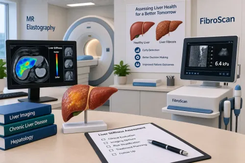
Elastography measures liver stiffness using imaging technology.
Vibration-controlled transient elastography is one commonly used method. It can provide a liver stiffness measurement without inserting a needle into the liver.
Magnetic resonance elastography is another option and can be particularly useful when more detailed non-invasive assessment is needed. AASLD notes that imaging-based techniques such as VCTE and MRE are established tools for evaluating fibrosis.
Can non-invasive testing replace biopsy?
Sometimes, but not universally.
The decision depends on what the doctor needs to know.
Non-invasive assessment can be highly useful when the main question is whether significant or advanced fibrosis is likely. In many patients, a combination of blood-based assessment and elastography can provide meaningful risk stratification.
Recent AASLD reporting on MASLD research found that vibration-controlled transient elastography performed similarly to histology for predicting major liver-related outcomes in a large study population.
However, biopsy may still have a role when:
- The diagnosis remains unclear
- Different liver diseases are being considered
- Non-invasive results conflict
- The exact histological pattern will influence management
- There is significant uncertainty despite imaging and laboratory assessment
Why one test should not be chosen automatically
A common mistake is assuming that the most advanced test is always the best test.
For some patients, a basic blood-based assessment may be a sensible starting point. Others may need elastography. Some may require more advanced imaging or biopsy.
The right sequence is usually determined by clinical risk rather than by convenience alone.
What patients should bring to a fibrosis consultation
If you are being evaluated in Mumbai for fatty liver or another chronic liver condition, bring previous:
- Liver function tests
- Complete blood counts
- Ultrasound reports
- Elastography reports
- Viral hepatitis results
- Diabetes and lipid reports
- Previous biopsy reports
- Medication and supplement lists
This allows the hepatologist to interpret the current findings alongside the patient's history.
For patients consulting Dr. Chetan Kalal Hepatologist and Liver Transplant Specialist, the discussion can focus not only on whether fibrosis is present but also on what the result means for future monitoring and treatment.
Non-invasive fibrosis assessment has changed the patient experience by making risk stratification possible without automatically proceeding to an invasive procedure. The key is using the right test for the right clinical question.
Keywords