Varices form when increased pressure in the portal venous system reroutes blood through fragile veins in the esophagus or stomach. By understanding the difference between screening and emergency treatment, you can see when endoscopy is useful, which procedure fits the varix, and when medicines or radiological treatment are needed instead.
Key takeaways
- Varices become dangerous when enlarged veins rupture and cause gastrointestinal bleeding.
- Endoscopy screens for varices and can treat bleeding during the same procedure.
- Esophageal varices often need band ligation; gastric varices require different techniques.
- Prevent another bleed with liver care, prescribed medicines, and follow-up endoscopy.
How Portal Hypertension Creates Varices—and When They Become Dangerous
Portal hypertension is abnormally high pressure in the portal vein, which carries blood from the digestive organs to the liver. In cirrhosis, scar tissue restricts blood flow through the liver, so blood is diverted into smaller veins along the lower esophagus and stomach. These veins enlarge, stretch, and become varices.
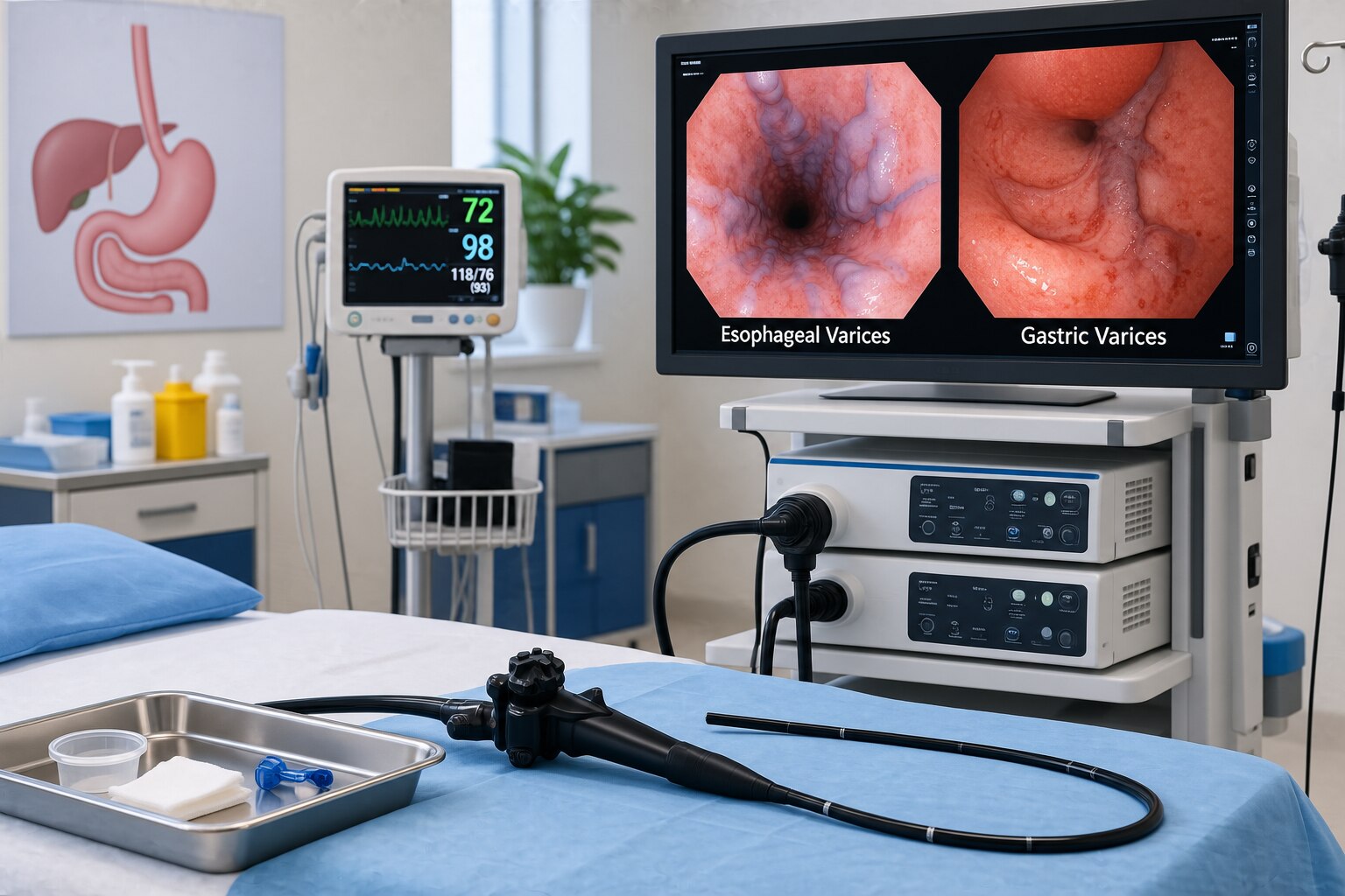
Esophageal varices are swollen veins in the esophageal wall. Gastric varices form in the stomach, often near the upper stomach or fundus. Their walls are thin and exposed to high pressure, so they can split and bleed heavily.
The danger rises with larger varices, red wale marks seen during endoscopy, advanced liver dysfunction, high portal pressure, or a previous variceal bleed.
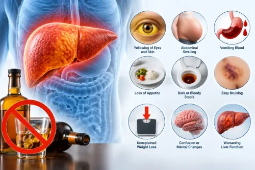
A rupture can cause vomiting of fresh blood or coffee-ground material, black tarry stools, weakness, fainting, or shock. Treat it as an emergency; do not wait for bleeding to stop.
Not every varix found during an endoscopy needs immediate treatment. Doctors weigh active bleeding, varix size and appearance, liver disease severity, and its location before choosing surveillance or preventive therapy.
Varices are also different from portal-hypertensive gastropathy, which creates a mosaic or snake-skin pattern and can ooze diffusely; banding a visible varix does not treat it. Endoscopy controls the bleeding site, not portal hypertension, so varices can recur or new ones can develop.
When Cirrhosis Calls for Screening or Therapeutic Endoscopy
Active vomiting of blood, black stools, dizziness, or low blood pressure makes endoscopy an emergency, not a screening appointment. After resuscitation, vasoactive medication and antibiotics, doctors usually perform upper endoscopy within 12 hours. Band ligation is preferred for active esophageal-variceal bleeding; gastric fundal varices often need cyanoacrylate injection or EUS-guided coil and glue treatment.
For a stable person with cirrhosis, the timing depends on bleeding history and risk:
- Previous variceal bleeding: arrange repeat therapeutic endoscopy as part of secondary prevention, usually with additional medication and scheduled banding until the varices are controlled.
- No previous bleeding but medium or large esophageal varices, or small varices with red marks or advanced liver disease: offer a nonselective beta blocker or prophylactic band ligation.
- No bleeding and only small, low-risk varices: do not band routinely; monitor with planned surveillance instead.
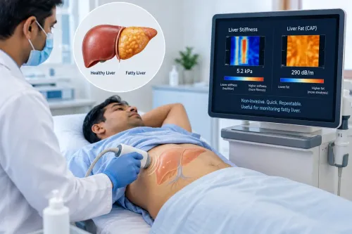
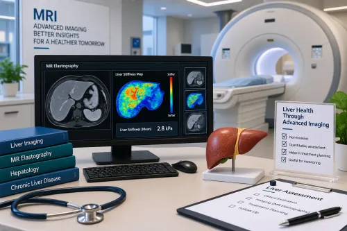
Doctors also use platelet count, liver stiffness, and imaging. A low platelet count, enlarged spleen, or collateral veins on ultrasound, CT, or MRI increases concern for clinically significant portal hypertension and can prompt endoscopy. Some patients with compensated disease and reassuring non-invasive measurements can safely delay it.
The scope must examine the esophagus, stomach, and gastroesophageal junction because gastroesophageal and isolated gastric varices require different treatment. Good esophageal varices endoscopy care includes urgent therapeutic capability, controlled sedation, and access to interventional radiology for TIPS or venous-obliteration procedures when endoscopy cannot control the problem.
What Happens During Varices Endoscopy Procedure and Esophageal Banding
A varices endoscopy procedure for suspected acute bleeding follows resuscitation, not replaces it. Doctors secure breathing and circulation, start a vasoactive drug such as terlipressin or octreotide, give preventive antibiotics, and perform endoscopy after stabilization, generally within 12 hours. You will usually fast and receive controlled sedation or anaesthesia.
- The endoscopist passes a flexible camera through your mouth to inspect the oesophagus, stomach, and duodenum, while suctioning blood or clots that obscure the view.
- For active oesophageal bleeding or high-risk oesophageal varices, the operator fits a transparent cap to the scope, suctions a varix into it, and releases a small elastic band around its base.
- The band cuts off blood flow. The trapped varix clots, shrinks, and later sloughs away, leaving scar tissue that lowers the chance of immediate bleeding from that spot.
- The team observes you during recovery and checks for recurrent bleeding, breathing problems, or unstable blood pressure before discharge or ward transfer.
A sore throat, chest discomfort, or difficulty swallowing can follow banding; black stools, vomiting blood, faintness, severe chest pain, or worsening breathlessness require urgent assessment. After a first bleed, one session rarely finishes treatment: repeat banding is commonly performed every 1–4 weeks until eradication, alongside a nonselective beta blocker when appropriate.
Rebleeding despite both treatments prompts consideration of TIPS or another specialist procedure. Finding varices alone does not automatically justify banding.
Why Gastric Varices Need a Different Endoscopic Approach
Band ligation works well for esophageal varices because an elastic band can strangulate the protruding vessel against the oesophageal wall. Endoscopy for gastric varices needs a different plan: fundal veins are larger, deeper, and connected to complex shunts, so bands can slip or fail to control blood flow.
Gastroesophageal varices type 1 (GOV1), which extend along the lesser curvature from oesophageal varices, may suit banding or glue. Fundal GOV2 and isolated gastric varices type 1 usually require glue-based treatment or interventional radiology.
| Option | What it does | When doctors consider it |
|---|---|---|
| Cyanoacrylate glue | Fills and seals the bleeding vein; EUS-guided coils with glue or another embolic material may improve targeting | Active bleeding fundal varices or suitable GOV1 anatomy |
| Vasoactive medicines | Reduce splanchnic blood flow and portal pressure; they support, rather than replace, definitive treatment | Start during suspected acute bleeding, alongside antibiotics and endoscopy |
| Balloon tamponade | Compresses bleeding temporarily | Rescue bridge when haemorrhage is uncontrolled; it is not a lasting treatment |
| BRTO | Blocks the draining vein through a balloon-occluded retrograde route | Fundal varices with a suitable gastrorenal or splenorenal shunt |
| TIPS or DIPS | Creates a portosystemic channel to lower portal pressure; DIPS accesses the portal vein directly through the liver | Refractory or recurrent bleeding, or anatomy unsuitable for BRTO or endotherapy |
The choice depends on CT or Doppler findings, the bleeding site, shunt anatomy, liver function, and local expertise. For gastric varices treatment in Mumbai, ask whether the service can provide urgent therapeutic endoscopy, controlled sedation, vasoactive drugs, antibiotics, and interventional radiology when glue alone is insufficient.
Choosing the Right Team and Preventing Another Variceal Bleed
A suitable unit must treat the cause of danger, not merely schedule a camera test. Ask whether it can provide urgent therapeutic endoscopy, vasoactive medication, antibiotics, monitored sedation or anesthesia, and rapid access to interventional radiology for TIPS or venous-obliteration procedures.
If you are arranging endoscopy for esophageal and gastric varices treatment in Mumbai, check these points before choosing a team:
- Can the team manage active bleeding, blood loss, and airway protection?
- Does it treat gastric varices with techniques beyond routine banding?
- Are hepatology, anesthesia, interventional radiology, and transplant services available when the first procedure is not enough?
- Who will arrange repeat endoscopy, medicines, and liver-disease follow-up?
A consultation with Dr Chetan Kalal Hepatologist Transplant should address the plan after discharge, not just the immediate procedure.
| Situation | What follow-up involves | Why it matters |
|---|---|---|
| Esophageal variceal bleed controlled | Repeat band ligation every 1–4 weeks until eradication, plus a nonselective beta blocker when appropriate | Varices can return before the next symptoms |
| Rebleeding despite both treatments | Assessment for early or rescue TIPS | Repeated endoscopy alone may not control portal pressure |
| Gastric varices or diffuse portal-hypertensive gastropathy | Specialist review of glue or venous-obliteration options, medicines, and portal-pressure reduction | Banding a visible varix does not treat mosaic “snake-skin” gastropathy |
Endoscopy closes the bleeding site; it does not remove portal hypertension. Ongoing liver treatment, surveillance, and assessment for TIPS or transplantation prevent a false sense of security.
Related services
Advanced Liver Procedures & Interventions" "Endoscopy for Esophageal and Gastric Varices Treatment in Mumbai, and India" Endoscopy for Esophageal & Gastric Varices Treatment in Mumbai (By Dr. View service → |
Emergency Upper GI Bleed Gastroenterology | Hepatology | GI Bleed Management Variceal Bleeding & Melena Treatment Malena & Variceal Bleeding: Life-Threatening GI Emergencies in Liver Disease Malena (black, tarry stools) and variceal bleeding are serious... View service → |
Frequently asked questions
When do cirrhosis patients need endoscopy for varices?
Patients with cirrhosis may need screening endoscopy based on their risk of clinically significant portal hypertension. Urgent endoscopy is required when vomiting blood, black stools, dizziness, or shock suggests bleeding.
What happens during an esophageal varices endoscopy procedure?
A flexible endoscope examines the esophagus and stomach. The doctor can place elastic bands around bleeding or high-risk esophageal varices to stop blood flow and reduce future bleeding.
How does gastric varices treatment differ from esophageal banding?
Gastric varices often need treatments such as tissue adhesive injection, endoscopic ultrasound-guided therapy, or radiological procedures because standard banding is not suitable for every gastric varix.
How can you prevent another variceal bleed?
Follow prescribed portal-pressure treatment, attend repeat endoscopy appointments, avoid alcohol, manage the underlying liver disease, and seek emergency care for any sign of gastrointestinal bleeding.
Keywords