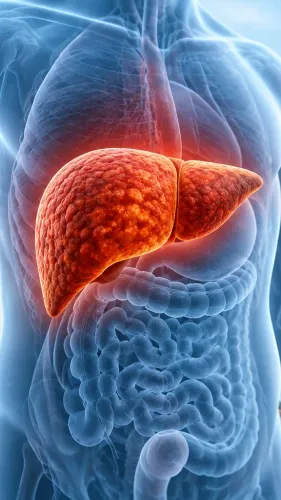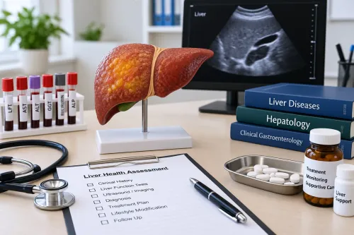
An MRE result is a measurement of how shear waves move through the liver, not a direct photograph of scar tissue. By comparing MRE with blood scores, ultrasound-based tests, FibroScan and biopsy, you can decide when its extra detail is worth the cost, time and MRI preparation.
Key takeaways
- MRE reports liver stiffness in kilopascals by tracking shear-wave movement.
- FibroScan is faster; MRE helps when results are uncertain or broader liver imaging is needed.
- Tell the imaging team about implants, pregnancy, claustrophobia, and recent food intake.
- Use the kPa result with blood tests, medical history, and imaging—not as a standalone diagnosis.
What does MR elastography actually measure?
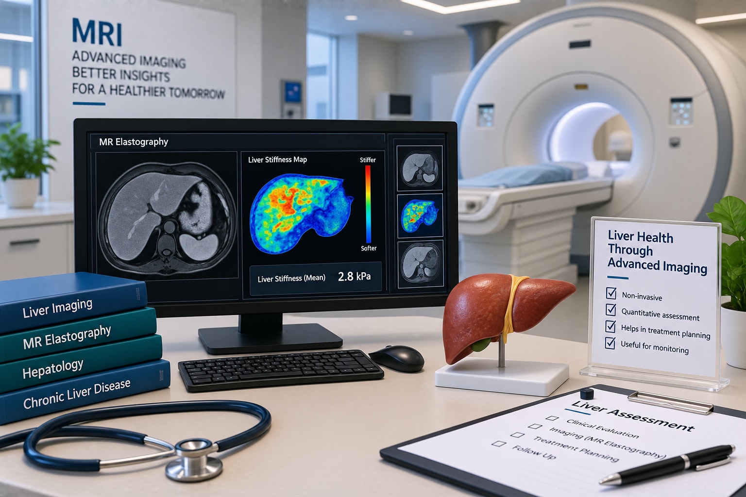
MR elastography (MRE) measures how shear waves travel through your liver, then reports that propagation as liver stiffness in kilopascals (kPa). An external vibration driver placed against the upper abdomen creates the waves; MRI records them and produces a liver stiffness measurement.
Stiffness is an indirect marker associated with fibrosis. It does not count scar tissue, show fibrosis under a microscope or provide a microscopic diagnosis. Acute hepatitis, steatohepatitis, bile-duct obstruction, heart-failure-related liver congestion and a recent meal can all raise stiffness without an equivalent amount of established fibrosis.
No universal kPa cutoff applies to every disease, MRI scanner or protocol. A value around 3.6 kPa is used in some specialist algorithms for suspected metabolic dysfunction-associated steatotic liver disease as a risk threshold, not as a biopsy-equivalent diagnosis. Interpret it with liver enzymes, bilirubin, platelets, imaging and symptoms.
A useful report should include:
- Technical adequacy, including whether motion or poor wave coverage limited interpretation
- A stiffness map showing the measured liver regions
- The result in kPa
- The MRI scanner details or MRE protocol when relevant
- The clinical reference range used
Compare follow-up values only when preparation and scanning technique are similar. A lower result can reflect less inflammation or congestion, not necessarily less fibrosis.
How does MRE compare with FibroScan and other non-invasive tests?
MRE provides a more reliable whole-liver stiffness assessment than FibroScan when results are unclear, discordant or technically unreliable. It is a non-invasive liver testing option and one of the important liver biopsy alternatives, without biopsy’s bleeding, pain, infection and sampling-error risks.
| Option | What it shows | Practical strengths and limits |
|---|---|---|
| FIB-4 and APRI | Blood-test-based fibrosis risk | Quick screening tools; they do not directly stage the whole liver |
| Laboratory panels and platelets | Liver injury, function and portal-hypertension clues | Add context but cannot map liver stiffness or confirm fibrosis stage |
| Ultrasound | Fat, portal-hypertension signs and structural abnormalities | Accessible, but it does not measure fibrosis reliably |
| Conventional MRI | Anatomy, fat, vessels and lesions | It measures stiffness only when a dedicated MRE sequence is added |
| FibroScan or ultrasound elastography | Local or limited-area stiffness | Faster, cheaper, portable and widely available; obesity, ascites, operator technique and limited sampling can distort results |
| MRE | Stiffness across a larger liver volume | Less often limited by obesity or ascites, but requires an MRI scanner, costs more, takes longer and may be difficult with claustrophobia or incompatible implants |
Choose MRE when FIB-4, APRI or FibroScan leaves fibrosis risk uncertain, or when test results conflict. It is not a universal biopsy replacement: MRE cannot identify the cause of injury, grade inflammation, confirm cancer or replace tissue when a treatment decision depends on microscopic diagnosis.
When is MRE the right next test—and when is it not?
MRE is the right next test when suspected fibrosis needs a more reliable whole-liver assessment and the result will change monitoring or referral. It fits unexplained ALT or AST elevation, fatty liver on ultrasound, hepatitis B or C, alcohol-related liver disease, an indeterminate FibroScan result, or suspected advanced fibrosis.
The clinician must read MRE beside bilirubin, liver enzymes, platelet count, metabolic status, imaging and symptoms. Acute inflammation, bile-duct obstruction or heart-failure-related congestion can raise stiffness without equivalent established fibrosis, so one kPa value cannot make the diagnosis.
Choose another immediate priority when you have:
- Urgent suspected liver failure, where rapid assessment of clotting, glucose, kidney function and complications comes first.
- Acute hepatitis, when inflammation can temporarily inflate liver stiffness.
- Severe abdominal pain requiring investigation for another diagnosis.
- An implanted device, metal fragment, severe claustrophobia or another reason you cannot safely undergo MRI.
MRE is one of the important liver biopsy alternatives, not a universal replacement. Biopsy can still be needed for suspected autoimmune, drug-induced, infiltrative or overlapping disease; strongly conflicting non-invasive results; or treatment and transplant decisions requiring tissue confirmation.
MRE alone cannot identify the cause of liver injury, grade inflammation, confirm liver cancer or determine transplant need. It is a form of non invasive liver testing that estimates risk; the wider assessment may still require blood tests, imaging, endoscopy or specialist review.
What should you disclose and prepare before an MRE appointment?
Before booking, give the imaging centre a complete MRI safety history. A device that is safe in one scanner or setting may not be safe in another.
1. Disclose a pacemaker or defibrillator, and provide the exact model of any MRI-conditional device. Also report implanted metal, a cochlear implant, retained metal fragments, an aneurysm clip, pregnancy or possible pregnancy, and severe claustrophobia.
2. Ask whether the centre can scan your specific device under its approved MRI conditions. Confirm that the appointment includes a dedicated liver MRE sequence, stiffness map and radiologist’s report—not only a routine abdominal MRI.
3. Confirm the fasting interval. Many departments request several hours without food because a recent meal can affect liver blood flow and stiffness. Ask how to handle diabetes medicines, insulin and regular medicines; do not change them without instructions.
4. Ask whether gadolinium contrast is planned. Stiffness measurement alone usually does not require contrast, but lesion assessment may. Tell staff about previous contrast reactions and kidney disease.
5. Wear clothing without metal. Remove jewellery, watches, bank cards, phones, hearing aids and other removable devices before entering the scanner room.
If you are arranging an mr elastography liver scan in mumbai, ask whether the final report states kPa units, technical adequacy and the scanner protocol or clinical reference range used. Bring prior scans and reports so the radiologist can compare technique and findings.
What happens during the scan, and how should you use the result?
During the scan, you lie on the MRI table with a firm external passive driver over your upper abdomen. The scanner makes loud knocking and humming sounds, so the centre provides ear protection. A technologist gives repeated short breath-hold instructions while the machine records vibration data. The examination takes longer than a quick ultrasound-based test.
Sedation is avoided when possible because you must follow those instructions. Discuss severe claustrophobia before booking.
Tell the imaging centre if you have:
- Trouble holding your breath
- A pacemaker, defibrillator or other implanted device
- Severe iron deposition
- A body size that could prevent safe positioning
Motion, poor breath-hold control, severe iron deposition or very large body size can make the study uninterpretable.
For an MR elastography liver scan in Mumbai, confirm that the appointment includes dedicated liver MRE, not only a routine abdominal MRI, and that the final report will contain a radiologist-reviewed stiffness map, liver stiffness measurement in kPa, technical adequacy and the reference range used.
| Result | How to use it | What to check |
|---|---|---|
| One measurement | Treat it as a fibrosis-risk marker, not a diagnosis | ALT, AST, platelets and disease cause |
| A changed serial value | Compare only with similar preparation and technique | Metabolic control, meals and scanner protocol |
| High stiffness with advanced disease | Arrange clinical review | Portal-hypertension signs, kidney function and decompensation symptoms |
Dr Chetan Kalal Hepatologist Transplant can place the result within decisions about monitoring, complications and transplant assessment; one number alone cannot determine treatment or transplant need.
Frequently asked questions
What does MR elastography measure?
MRE measures how shear waves travel through the liver and reports their propagation as liver stiffness in kilopascals (kPa).
How does MRE compare with FibroScan?
FibroScan is quicker and more accessible, while MRE examines a larger volume of liver and can be useful when FibroScan results are unreliable or unclear.
When is MRE the right next test?
MRE is useful when non-invasive tests disagree, FibroScan cannot produce a reliable reading, or your clinician needs liver stiffness alongside MRI assessment.
What should you disclose before an MRE appointment?
Tell the imaging team about metal implants, implanted electronic devices, pregnancy, claustrophobia, prior MRI problems, and recent food or drink.
How should you use an MRE result?
Interpret the kPa value with your blood tests, symptoms, medical history, and other imaging; do not use it alone to diagnose cirrhosis or determine treatment.
Keywords